 HUN-REN Biological Research Centre, Szeged




Biomembranes are the most fundamental and most abundant barriers of life. They are “smart” because their lipid bilayer serves as the structural basis whereas membrane-associated proteins regulate transfer of matter and information across this barrier, manifested as well-controlled biological processes. Our own biological subjects are ion-transporting membrane proteins, cellular plasma- and organelle membranes, and soluble proteins interacting with membranes or bio-mimetic surfaces, all with great relevance to human health and environment. Our overall approach is molecular biophysics as we use methods from Physics to address problems in Biology, down to molecular or even atomic details. We are keen on ever enhancing our experimental and theoretical techniques. Consequently, we have a wide palette of state-of-the-art spectroscopic and microscopic methods. The combination of the research infrastructure in our three laboratories (multimodal spectroscopy, femtosecond biology and atomic force microscopy) represents a synergy of complementary techniques that uniquely addresses biomolecular systems at high spatial, temporal and spectral resolution. As our research proved it, the high resolution data emerging from these techniques has sufficient information capacity to fingerprint distinct physiological states of biological samples. This molecular biophysics suite of methods also supports numerous external projects in our extensive network of in-house, national and international collaborations, all related to improving the quality of life.
Quick links
• A more detailed page of the Research Group.
• Full list of recent publications of the Research Group.
• The Integrated Paramagnetic Resonance and Optical Multi-Dimensional Bio-Spectroscopy (outstanding research infrastructure) and detailed list of research instruments.
• Popular science profile on facebook and videos on youtube.
• Our Research Topic about Ductin proteins and rotary ATPases @ Frontiers.
• Cover story of the Hungarian Academy of Sciences about our paper on the vacuolar proton-ATPase.
Laboratory of multi-modal spectroscopy of membranes and proteins
In this laboratory the main techniques are spin-labelling and spin-trapping electron paramagnetic resonance (EPR) [Csonka et al., 2015; Kóta et al., 2020; Marsh and Páli, 2004; Páli and Kóta, 2026] (Fig. 1), modular fluorescence [Böde et al., 2026; Sebők-Nagy et al., 2023a], infrared (FTIR) [Kóta et al., 2004; Kóta et al., 2020; Marsh and Páli, 2001; Páli and Marsh, 200] and fast kinetic absorption spectroscopies [Khoroshyy et al., 2021; Misra et al., 2023], calorimetry [Dér et al., 2007] and computational techniques [Bashtovyy et al., 2001, 2003; Koto et al., 2025; Krishnan et al. 2026; Marsh and Páli, 2001].
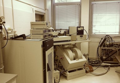
Figure 1. Our Bruker ELEXSYS E580 X-band CW and pulsed-, FT- electron paramagnetic resonance (EPR) spectrometer – the best, and most equipped state-of-the-art continuous wave and pulsed X-band EPR laboratory of Central- and East Europe.
The physical basis of protein function
The physical factors behind protein function are the electrostatic interaction between charges and dipoles; electron affinity and the related electron transfer reactions; proton affinity (pK) and the related proton transfer reactions as well as hydrogen bonds; steric interactions depending on the 3D protein structure; as well as the interactions with the water molecules inside and around the protein and with the surrounding lipid molecules in case of the membrane proteins. For proteins with chromophoric prosthetic groups, spectroscopic and kinetic spectroscopic studies in the visible-UV range (Fig. 2) provide essential information about the protein function. We study the photocycle and the molecular steps of the proton transport in bacterial retinal protein proton pumps, e.g. bacteriorhodopsin, or in signaling proteins, e.g. PYP, and their dependence on environmental factors. We investigate the structural factors affecting the kinetics of electron transfer in cytochromes. We develop and apply general chemometric methods for the analysis of spectral data matrices in order to determine the underlying physical phenomena. In addition, we study the possibilities of the integration of „colored” proteins in biophotonic and in bioelectronic applications. Our main experimental setup consists of a tunable Nd-Yag + OPO pulse laser to provide < 10 ns actinic pulses and a continuous white measuring light source in the entire visible range. Time resolved spectra of the excited sample can be measured at delay times from < 100 ns upwards. High time resolution single wavelength kinetics can also be measured in the same experimental arrangement.
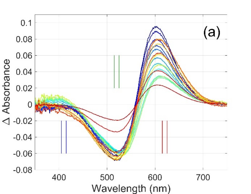
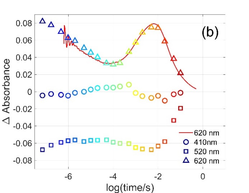
Figure 2. Multiwavelength and single wavelength absorption change signals measured on Gloeobacter rhodopsin [Misra et al., 2023]. Symbols in (b) are the amplitudes obtained at the selected wavelengths from the difference spectra in (a).
Membrane-associated proteins of pH regulation and membrane fusion processes of organelles and transport vesicles
The molecular mechanisms of pH regulation and membrane fusion of the eukaryotic cellular organelles are of high relevance to many normal and pathogenic physiological processes. We focus on 3 classes of key proteins in these processes: the vacuolar proton-ATPase (V-ATPase), the Ductin and Syntaxin proteins. The former two are strongly related since part of the V-ATPase belongs to the Ductin family. The protein complex that is responsible for the acidification of internal organelles of eukaryotic cells is nature's most universal proton pump, the vacuolar proton-ATPase [Abuammar et al., 2021]. V-ATPase also takes part in membrane fusion processes and plays crucial roles in a number diseases, and its tissue-specific inhibition is a key therapeutic objective. V-ATPase is a membrane-bound molecular rotary engine. We were the first to report the rotation rate in a V-ATPase that was not subjected to genetic or chemical modification and also not fixed to a solid support [Ferencz et al., 2013], and also discovered that the activity of V-ATPase can be affected with an oscillating trans-membrane electric field [Ferencz et al., 2017; Petrovszki et al., 2021] (Fig. 3). Ductins and syntaxins play important roles in vesicle fusion mechanisms. Since our discovery of the Ductin family [Holzenburg et al., 1993], we have determined the membrane assembly and interaction of one of the gap-junction Ductin proteins with lipids [Kota et al., 2008; Pali et al., 1995; Pali et al., 1999]. We have recently identified new structural features of the Ductin family and potential new factors regulating their membrane assembly [Sebők-Nagy et al., 2023b]. Our current studies aim at the mutual effects of V-ATPase and its molecular interface, including the effect of synthetic inhibitors and divalent cations on the enzymatic activity, subunit assembly and the rotary mechanism [Páli et al., 2024]. In collaboration with the Drosophila Autophagy group (Institute of Genetics) we are studying the binding of Syntaxins to membranes [Laczkó-Dobos et al., 2024] and search for new factors regulating lysosome acidification through V-ATPase.
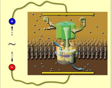
Figure 3. The ATP consumption of a native V-ATPase can be altered by an oscillating trans-membrane potential, and the effect is dependent on the relationship between the main frequencies in the stimulation and the rate of rotation in the enzyme. (Taken from [Petrovszki et al., 2021].)
Folding, insertion, structure and assembly of proteins in the membrane and at the membrane-water interface
An experimentally and computationally challenging problem in biophysics is how membrane proteins are inserted, folded and assembled in the lipid bilayer, in some cases even pass it through, often in the absence of folding assistants and take up their native biologically functional conformation. In this membrane-protein folding problem specific interactions between the protein and lipids of the target membrane play a crucial role yet to be explored in detail. In reconstituted lysozyme-lipid complexes, we are studying the influence of the lipid bilayer on activity, thermal (un)folding and stability of this antibacterial enzyme. Since even closely related soluble proteins unfold or refold at the membrane-water interface very differently, it is an interesting phenomenon to be understood. In the past, we have modelled the membrane-embedded structure of several membrane proteins [Bashtovyy et al., 2003; Pali et al., 2006; Kostrzewa et al., 2000] and polypeptides [Bashtovyy et al., 2001; Kota et al., 2004; Kota et al., 2008] based on spectroscopic structural data. Currently we are focusing on a more systematic combination of experimental and theoretical techniques, including machine learning, for structural biology of membrane proteins. Our approach is to measure structural data on the target biomolecular complexes in their functional state and native environment, and combine the so-obtained data with theoretical methods in order to obtain the 3-dimensional fold or complete complex systems (Fig. 4, [Koto et al., 2026]).
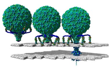
Figure 4. Our entry in the “gigastructures” database [Koto et al., 2026] represents the first atomic structure of a complete bacteriophage, namely the T7 phage of E.coli. The image shows the main stages of phage entry.
Protein interfaces and structural reorganisations in natural and synthetic membranes and lipid-based drug delivery formulas
Uncoupling or perturbing the selective structural and dynamic balance between membrane proteins and lipids may either force the organism to adapt to the new conditions, by e.g. modifying its lipid composition, or die when unable to do so. The protein-lipid interface takes several different forms, all of which are crucial to biology. For biological membranes the protein-lipid interface may be either polar in the case of surface-bound or absorbed peripheral proteins, or apolar in the case of integral transmembrane proteins. In collaborations with the Institute of Plant Biology, we are studying the role of non-bilayer lipid phases and free radicals [Kota et al., 2002; Lingvay et al., 2020; Rehman et al., 2021] in adaptive reorganisations and stress responses, respectively, of photosynthetic membranes. In domestic and international collaborations we are studying the membrane dynamics and lipid-protein interaction in lipid-based drug delivery formulas [DeNardis et al., 2026; Klacsová et al., 2026] and the effect of waste-water TiO2-based filtration membranes on soluble proteins [Sebők-Nagy et al., 2023a] (Fig. 5) and characterise nano metal-organic frameworks membranes for purification of waste water from organic contaminates [Emam et al., 2024], respectively.
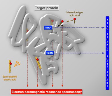
Figure 5. The graphical abstract of our recent spectroscopic study of changes in the structure and dynamics of bovine serum albumin exposed to TiO2 nanoparticles or passing through commercial polyvinylidene fluoride membranes coated with the same nanoparticles [Sebők-Nagy et al, 2023a].
Femtobiology laboratory
Fluorescence spectroscopy with femtosecond time resolution
A measuring system suitable for the detection of fluorescence kinetics in the 100 fs – 10 ns time interval and in the complete visible spectral range was constructed [Khakurel et al., 2025]. Using this apparatus we are studying the coenzymes FAD and NADH – substantial components of biological redox systems – in different microenvironments. We have found that in aqueous solution the kinetics of FAD fluorescence (Fig. 6) can be characterized by several time constants: one of them, falling in the ns range, can be associated to the open conformation of the molecule, while other components in the ps range correspond to closed conformations. The Hofmeister effect – controlling the ratio of molecules in the different conformational states – is well observable on the kinetics, in accordance with the models based on molecular dynamics calculations. The setup also enables the observation of the Stokes-shift, characterized by a lifetime of ~100 fs, and the study of the related solvation dynamics. In contrast to the above parameters, in the case of FAD bound covalently to the flavocytochrome c sulfide dehydrogenase enzyme, we observed only a single sub-ps component, indicating that the coenzyme is in an extremely strong interaction with the protein microenvironment. Regarding NADH the equilibrium of the open and closed conformations is also traceable, however, the Stokes-shift has three components, related to an unusually complex process of vibrational relaxation.
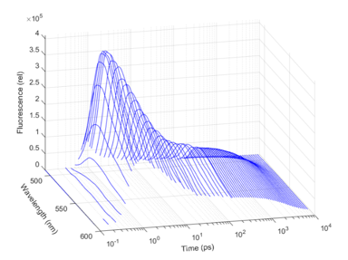
Figure 6. Fluorescence kinetics of FAD in time and wavelength domain
The above-mentioned reactions, taking place in the excited state, can be characterized by first-order kinetics, similarly to the light-induced ground-state reactions of retinal proteins. The standard way of the analysis of such complex system reactions is based on the method of nonlinear regression with exponential terms, whose application faces many difficulties. The major problem is that small noise on the experimental data generates high uncertainties in the predicted parameters, and even in the number of the necessary parameters. Using simulated data based on a very complex model of the bacteriorhodopsin photocycle we elaborated a new machine learning method for handling this problem [Zimányi et al., 2021] by applying techniques of modern statistics (cross-validation, Bayesian optimization), which performs very well on the analysis of the experimentally detected fluorescence kinetics as well.
A multifunctional femtobiology workstation
Based on the unique infrastructure of the ELI ALPS Research Institute in Szeged, collaborating with groups of the HUN-REN BRC and the University of Pécs, we are developing a permanent workstation with the purpose of studying ultrafast processes on biological systems utilizing light energy (light-harvesting complexes and photosynthetic reaction centers, retinal proteins, plant photoreceptors) [Akhtar et al., 2025]. The measuring techniques are founded partially on the expansion of the most modern technology of femtosecond multidimensional electronic spectroscopy [Singhal et al., 2025], and partially on the improvement of the methods we developed for the detection of low-intensity light-induced coherent THz radiation.
AFM and Raman microscopy laboratory
Biological applications of scanning probe and confocal Raman microscopy
The laboratory performs structural, morphological and mechanical investigation of biological objects, ranging from single protein molecules up to (the plasma membrane of) living cells and tissues. Nanometer scale mechanical manipulation and measurement in a liquid environment provides an absolute advantage compared to conventional cellular imaging techniques. The wide operation scale offers investigation of pico-Newton range intermolecular forces up to nano-Newton intercellular forces, even micro-Newton cell to substrate forces. Towards the understanding and the description of tumors and metastasis formation, mechanical investigations have recently gained increasing interest [Varga et al., 2017]. The ability to directly investigate the interaction of tumor cells with their environment offers outstanding opportunities. One of the active topics of the laboratory is the investigation of the mechanical interaction between tumor cells and brain endothelial cells or pericytes [Csonti et al., 2024; Fazakas et al., 2021]. Figure 7 depicts a reconstructed force-coloured adhesion map recorded with a tumor cell as a probe.
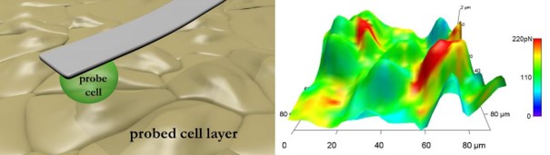
Figure 7. Left: schematic representation of the measurement setup. Right: adhesion force-colored height reconstruction of endothelial cells recorded with a living melanoma cell as a probe.
Atomic force microscopy augmented by label-free chemical imaging based on Raman scattering (Fig. 8) is a very potent physicochemical technique also for biological material. The method has emerged as a non-destructive and non-invasive tool to intracellularly detect administered drugs without exogenous labelling. Among the most promising applications one can find the ability of discrimination between normal and tumor cells not only in vitro, but in vivo as well. Co-localized topography, stiffness and chemical maps offer outstanding opportunities to investigate structural and dynamic processes. Successful projects included self-assembly of oligonucleotides [Bálint et al., 2007], structural and morphological studies of nano and microvesicles [Laczkó-Dobos et al., 2021], mechanical investigation of different bacterial strains [Miclea et al., 2010], plasticity of red blood cells [Chakraborty et al. 2019], de-adhesion patterns of living brain endothelial cells [Csonti et al., 2024] and quantitative mapping of several tissues. The above list is far from being complete, it only serves the presentation of the large scale and subject areas where our system can provide valuable information.
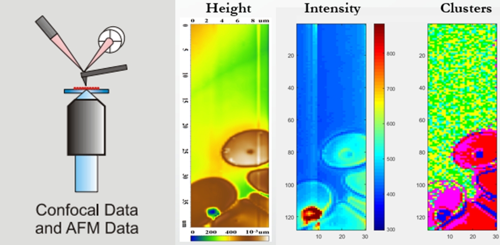
Figure 8. Scheme of the device combining confocal Raman spectroscopy and atomic force microscopy. Combined topography, Raman scattering intensity and spectral discriminative mapping of red blood cells.
Selected publications of the research group
Abuammar, H., Bhattacharjee, A., Simon-Vecsei, Zs., Blastyak, A., Csordas, G., Pali, T. and Juhasz, G. (2021) Ion Channels and Pumps in Autophagy: A Reciprocal Relationship. Cells 2021, 10(12), 3537.
Akhtar, P., Wang, W., Singhal, S., Sarlós, F., Shen, J-R. and Lambrev, P. (2025) Excitation energy transfer and trapping in Photosystem I of the green alga Bryopsis corticulans. Photosynthesis Research 163, 62.
Bálint, Z., Nagy, K., Laczkó, I., Bottka, S., Végh, G.A., Szegletes, Z. and Váró, G. (2007) Adsorption and Self-Assembly of Oligodeoxynucleotides onto a Mica Surface.J. Phys. Chem. C 111(45), 17032–17037.
Bashtovyy, D., Marsh, D., Hemminga, M.A. and Páli, T. (2001) Constrained modeling of spin-labeled major coat protein mutants from M13 bacteriophage in a phospholipid bilayer. Protein Sci 10, 979-987.
Bashtovyy, D., Berczi, A., Asard, H. and Pali, T. (2003) Structure prediction for the di-heme cytochrome b(561) protein family. Protoplasma 221, 31-40.
Bérczi, A., Márton, Z., Laskay, K., Tóth, A., Rákhely, G., Duzs, Á., Sebők-Nagy, K., Páli, T. and Zimányi, L. (2023) Spectral and redox properties of a recombinant mouse cytochrome b561 protein suggest transmembrane electron transfer function. Molecules 28(5):2261.
Böde, K., Sebők-Nagy, K., Dlouhy, O., Steinbach, G., Benda, A., Garab, Gy. and Páli, T. (2026) Lipid assemblies mediating the fusion of photosystem-II enriched membranes characterized by DPH (1,6-diphenyl-1,3,5-hexatriene) fluorescence and fluorescence anisotropy lifetimes. Journal of Photochemistry and Photobiology B 275, 113361.
Chakraborty, P., Krisztina N., Dugmonits, K.N, Végh, A.G., Hollandi, R., Horváth, P., Maléth, J., Hegyi, P., Németh, G. and Hermesz, E. (2019) Failure in the compensatory mechanism in red blood cells due to sustained smoking during pregnancy.Chemico-Biological Interactions 313(1) 108821.
Csonka, C., Pali, T., Bencsik, P., Gorbe, A., Ferdinandy, P. and Csont, T. (2015) Measurement of NO in biological samples. Br J Pharmacol 172, 1620-1632.
Csonti, K., Fazakas, C., Molnár, K., Wilhelm, I., Krizbai, I.A., and Végh, A.G. (2024) Breast adenocarcinoma cells adhere stronger to brain pericytes than to endothelial cells. Coll. And Surf. B Biointerfaces 234, 113751.
DeNardis, N.I., Radić, T.M., Klacsová, M., Uhrikova, D., Frkanec, R., Zemła, J., Lekka, M., Lukeš, M., Prasil, O., Sebők-Nagy, K., Pali, T., Lisac, K., Chovancová, M., Ojdanic, K., Horvat, L., Štefanić, P.P., Smokrović, K., Maltar-Strmečki, N., Saftić, D.P., Piantanida, I. and Glavan, T.M. (2026) Biophysical approach to structural characterization of biocompatible microalgae-derived vesicles as sustainable drug carriers. Algal Research 104852.
Dér, A., Kelemen, L., Fabian, L., Taneva, S.G., Fodor, E., Pali, T., Cupane, A., Cacace, M.G. and Ramsden, J.J. (2007) Interfacial water structure controls protein conformation. J Phys Chem B 111, 5344-5350.
Emam, H.E., Koto, T., Sebok-Nagy, K., El-Shahat, M., Abdel-Gawad, H., Pali, T. and Abdelhameed, R.M. (2024) Synthesis, spectroscopic study and carbofuran adsorption of mixed metal (Co, Cu)@Ca-BTC frameworks aimed at wastewater cleaning. Journal of Industrial and Engineering Chemistry 139, 444-457.
Fazakas, C., Kozma, M., Molnár, K., Kincses, A., Dér, A., Fejér, A., Horváth, B., Wilhelm, I., Krizbai, I.A., and Végh, A.G. (2021) Breast adenocarcinoma-derived exosomes lower first-contact de-adhesion strength of adenocarcinoma cells to brain endothelial layer. Colloids Surf. B Biointerfaces 204, 111810.
Ferencz, C., Petrovszki, P., Kota, Z., Fodor-Ayaydin, E., Haracska, L., Bota, A., Varga, Z., Der, A., Marsh, D. and Pali, T. (2013) Estimating the rotation rate in the vacuolar proton-ATPase in native yeast vacuolar membranes. European Biophysics Journal 42, 147–158.
Ferencz, Cs-M., Petrovszki, P., Der, A., Sebok-Nagy, K., Kota, Z. and Pali, T. (2017) Oscillating electric field measures the rotation rate in a native rotary enzyme. Scientific Reports 7, 45309.
Holzenburg, A., Jones, P.C., Franklin, T., Páli, T., Heimburg, T., Marsh, D., Findlay, J.B.C, Finbow, M.E (1993) Evidence for a common structure for a class of membrane channels. Eur J Biochem 213(1) 21-30.
Khakurel, K.P., Fuertes, G., Sipos, A., Paragi, G., Dostal, J., Kloz, M., Žoldák, G., Andreasson, J. and Tóth, A. (2025) Ultrafast Dynamics in Flavocytochrome C by Using Transient Absorption and Femtosecond Fluorescence Lifetime Spectroscopy. The Journal of Physical Chemistry B 129(15), 3731-3739.
Khoroshyy, P., Tenger, K., Chertkova, R.V., Bocharova, O.V., Kirpichnikov, M.P., Borovok, N., Groma, G.I., Dolgikh, D.A., Kotlyar, A.B. and Zimányi, L. (2021) Kinetics and energetics of intramolecular electron transfer in single-point labeled TUPS-cytochrome c derivatives. Molecules 26(22):6976.
Klacsová, M., Sebők-Nagy, K., Páli, T., Chovancová, M., Almásy, L. and Uhríková, D. (2026) Microalgae-derived vesicles as potential carriers for therapeutic biomacromolecules. Journal of Drug Delivery Science and Technology 117, 107985.
Kostrzewa, A., Pali, T., Froncisz, W. and Marsh, D. (2000) Membrane location of spin-labeled cytochrome c determined by paramagnetic relaxation agents. Biochemistry 39(20), 6066-6074.
Kóta, Z., Horvath, L.I., Droppa, M., Horvath, G., Farkas, T. and Pali, T. (2002) Protein assembly and heat stability in developing thylakoid membranes during greening. Proceedings of the National Academy of Sciences USA 99, 12149-12154
Kóta, Z., Páli, T. and Marsh, D. (2004) Orientation and lipid-peptide interactions of gramicidin A in lipid membranes: polarized attenuated total reflection infrared spectroscopy and spin-label electron spin resonance. Biophys J 86, 1521-1531.
Kóta, Z., Pali, T., Dixon, N., Kee, T.P., Harrison, M.A., Findlay, J.B.C., Finbow, M.E. and Marsh, D. (2008) Incorporation of transmembrane peptides from the vacuolar H+-ATPase in phospholipid membranes: spin-label electron paramagnetic resonance and polarized infrared spectroscopy. Biochemistry 47(12), 3937–3949.
Koto, T., Ozvoldik, K. and Páli, T. (2026) Model of the T7 bacteriophage entry states. Version 26.3.20 , http://download.yasara.org/petworld/t7phageentry/index.html . Accession code: t7phageentry Accession code: t7phage @ Database: A database of biomolecular models with millions to billions of atoms for coarse-grained MD simulation and all-atom visualization ( http://download.yasara.org/petworld/index.html ).
Krekic, S., Zakar, T., Gombos, Z., Valkai, S., Mero, M., Zimányi, L., Heiner, Z. and Dér, A. (2020) Non-linear optical investigation of microbial chromoproteins. Front. Plant Sci 11:547818.
Krishnan, S.B., Winfred, J.S.R.V., Hilinski, E.F., Saltiel, J. and Zimányi, L. (2026) Transient absorption spectroscopy of the stilbenes in methanol – adiabatic cis -> trans photoisomerization by sequential two-photon excitation and revised ether quantum yields. J. Phys. Chem. A 130(37): 7404–7411.
Laczkó-Dobos H., Maddali, A.K, Jipa A., Bhattacharjee, A., Végh, A.G., Juhász, G. (2021) Lipid profiles of autophagic structures isolated from wild type and Atg2 mutant Drosophila. BBA - Molecular and Cell Biology of Lipids 1866(3), 158868.
Laczkó-Dobos, H., Bhattacharjee, A., Maddali, A.K., Kincses, A., Abuammar, H., Sebők-Nagy, K., Páli, T., Dér, A., Hegedűs, T., Csordás, G. and Juhász, G. (2024) PtdIns4p is required for the autophagosomal recruitment of STX17 (syntaxin 17) to promote lysosomal fusion. Autophagy 20 (7).
Lingvay, M., Akhtar, P., Sebok–Nagy, K., Pali, T. and Lambrev, P.H. (2020) Photobleaching of Chlorophyll in Light-Harvesting Complex II Increases in Lipid Environment. Frontiers in Plant Science 11, Article 849.
Marsh, D. and Páli, T. (2001) Infrared dichroism from the X-ray structure of bacteriorhodopsin. Biophys J 80, 305-312.
Marsh, D. and Pali, T. (2004) The protein-lipid interface: perspectives from magnetic resonance and crystal structures. Biochimica et Biophysica Acta 1666, 118-141.
Miclea, P.S, Péter, M., Végh, G., Cinege, G., Kiss, E., Váró, G., Horváth, I. and Dusha, I. (2010) Atypical Transcriptional Regulation and Role of a New Toxin-Antitoxin-Like Module and Its Effect on the Lipid Composition of Bradyrhizobium japonicum. Molecular Plant-Microbe Interactions 23(5), 638-650.
Misra, R., Das, I., Dér, A., Steinbach, G., Shim, J-G., Busse, W., Jung, K-H., Zimányi, L. and Sheves, M. (2023) Impact of protein-chromophore interaction on the retinal excited state and photocycle of Gloeobacter rhodopsin: role of conserved tryptophan residues. Chemical Science 14:9951.
Páli, T., Finbow, M.E., Holzenburg, A., Findlay, J.B.C., and Marsh, D. (1995) Lipid-Protein Interactions and Assembly of the 16-kDa Channel Polypeptide from Nephrops norvegicus. Studies with Spin-Label Electron Spin Resonance Spectroscopy and Electron Microscopy. Biochemistry 34(28), 9211-9218.
Páli, T., Finbow, E.M. and Marsh, D. (1999) Membrane assembly of the 16-kDa proteolipid channel from Nephrops norvegicus studied by relaxation enhancements in spin-label ESR. Biochemistry 38(43), 14311-14319.
Páli, T. and Marsh, D. (2001) Tilt, Twist, and Coiling in β-Barrel Membrane Proteins: Relation to Infrared Dichroism. Biophysical Journal 80, 2789-2797.
Páli, T., Bashtovyy, D. and Marsh, D. (2006) Stoichiometry of lipid interactions with transmembrane proteins - deduced from the 3-D structures. Protein Science 15(5), 1153-1161.
Páli, T. and Kóta, Z. (2026) Electron paramagnetic resonance spectroscopy of spin-labeled lipids for studying lipid-protein interaction in model- and biomembranes. Methods in molecular biology 3001, 555-588.
Páli, T., Feniouk, B. and Wilkens, S. (2024) Editorial: Functions, Working Mechanisms, and Regulation of Rotary ATPases and Ductin Proteins. Frontiers in Molecular Biosciences 11, 1399421.
Petrovszki, P., Sebők-Nagy, K. and Páli, T. (2021) The activity of native vacuolar proton-ATPase in an oscillating electric field – Demystifying an apparent effect of music on a biomolecule Frontiers in Molecular Biosciences 8, 772167.
Petrovszki, D., Valkai, S., Kelemen, L., Nagy, L., Agarwal, V., Krekic, Sz., Zimányi, L. and Dér, A. (2023) Microsecond all-optical modulation by biofunctionalized porous silicon microcavity. Nanomaterials 13:2070.
Rehman, A.u., Bashir, F., Ayaydin, F., Kota, Z., Pali, T., Vass, I. (2021) Proline is a quencher of singlet oxygen and superoxide both in vitro and in plant systems. Physiologia Plantarum 172, 7–18.
Sebők-Nagy, K., Kóta, Z., Kincses, A., Fazekas, Á.F., Dér, A., László, Z. and Páli, T. (2023a) Spin-Label Electron Paramagnetic Resonance Spectroscopy Reveals Effects of Wastewater Filter Membrane Coated with Titanium Dioxide Nanoparticles on Bovine Serum Albumin. Molecules 28(19), 6750.
Sebők-Nagy, K., Blastyák, A., Juhász, G. and Páli, T. (2023b) Reversible binding of divalent cations to Ductin protein assemblies-a putative new regulatory mechanism of membrane traffic processes. Frontiers in Molecular Biosciences 10, 1195010.
Singhal, S., Akhtar, P., Sarlós, F., Sipka, G., Sipos, Á., Jójárt, P., Gilicze, B., Seres, I., Bengery, Z., Tan, H. and Lambrev, P. (2025) Fast-scan pulse-shaper-assisted two-dimensional electronic spectroscopy of Photosystem I using high-repetition-rate supercontinuum pulses. Optics Express 33(26), 53851-53866.
Varga, B., Fazakas, C., Molnár, J., Wilhelm, I., Domokos, R.A., Krizbai, I.A., Szegletes, Z., Váró, G., and Végh, A.G. (2017) Direct mapping of melanoma cell - endothelial cell interactions. J. Mol. Recognit. 30(6):e2603.
Zimányi, L., Sipos, Á., Sarlós, F., Nagypál, R. and Groma, G. (2021) Machine learning-based model selection and parameter estimation from kinetic data of complex first-order reaction systems. PLOS One 16(8): e0255675.
Zimányi, L., Thekkan, S., Eckert, B., Condren, A.R., Dmitrenko,O., Kuhn, L.R., Alabugin, I.V. and Saltiel, J. (2020) Determination of the pKa values of trans-Resveratrol, a Triphenolic Stilbene, by Singular Value Decomposition. Comparison with Theory. J. Phys. Chem. A 124(31):6294-6302.
scientific adviser

research professor emeritus

research fellow

research fellow

research fellow

research fellow

research fellow

research fellow

scientific administrator
laboratory assistant

PhD student

PhD student

MSc student

BSc student

BSc student

BSc student
|
Tibor PÁLI
|
scientific adviser | publications | CV |
|
László ZIMÁNYI
|
research professor emeritus | publications | CV |
|
Krisztina SEBŐKNÉ NAGY
|
research fellow | publications | CV |
|
Áron SIPOS
|
research fellow | publications | CV |
|
Attila Gergely VÉGH
|
research fellow | publications | CV |
|
Teruaki KOTO
|
research fellow | publications | CV |
|
Zoltán KÓTA
|
research fellow | publications | CV |
|
András TÓTH
|
research fellow | publications | CV |
|
Ferenc SARLÓS
|
scientific administrator | publications | CV |
|
Csilla BARTA
|
laboratory assistant | publications | CV |
|
Katalin CSÁNYI
|
PhD student | publications | CV |
|
Sakina MIR
|
PhD student | publications | CV |
|
Réka BOLEGA
|
MSc student | publications | CV |
|
Richárd JANKÓ
|
BSc student | publications | CV |
|
Péter PASKA
|
BSc student | publications | CV |
|
Noémi SINYI
|
BSc student | publications | CV |
 back