 HUN-REN Biological Research Centre, Szeged

About us
The Cellular Imaging Laboratory provides (fluorescence) imaging services for HUN-REN BRC researchers, for collaborators from the SZTE and other Hungarian institutes, and for EuroBioImaging members. The available equipment consists of confocal laser scanning microscopes, fluorescence and stereo microscopes. Our expertise, image processing software collection and the available courses are supporting the efficient research.
Reservation system for the microscopes
Expertise and equipment
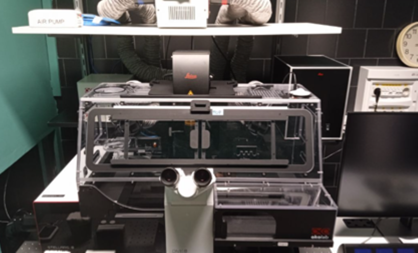
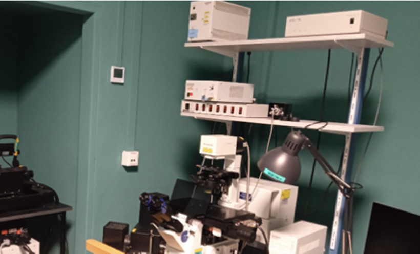
Leica Stellaris 8 WLL + FALCON
Our Leica Stellaris 8 Confocal Miscroscope is suitable for transmission imaging (with laser scanning, non-confocal), bright field illumination and fluorescence imaging (DAPI, FITC, FITC LP, TXR cubes) with camera, 6 objectives (10x/0.3, 20x/0.4, 20x/0.75, 40x/0.8, 40x/1.25 GLYC and 63x/1.4 OIL), confocal fluorescence imaging modes (Tau sense and FALCON) are available from 440 to 790 nm excitation. Long term measurements are supported by the Adaptive Focus Control and an Okolab Incubator (temperature, CO2).
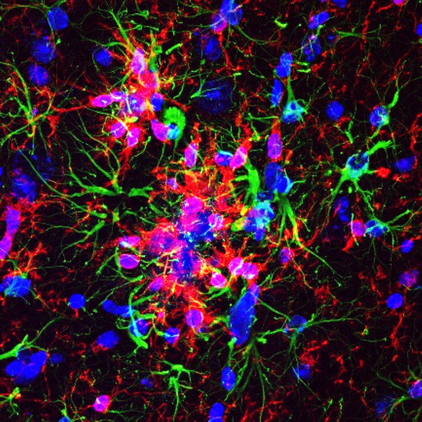

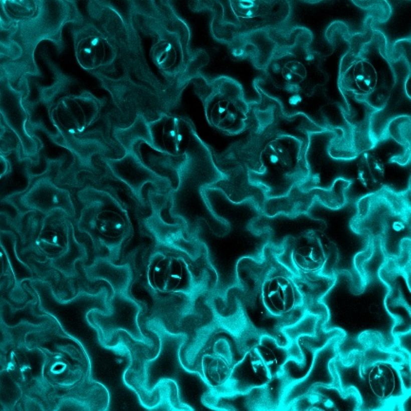
Microscopic photos by: Rákóczi Bettina & Nusser Zsombor; Diósi Ákos
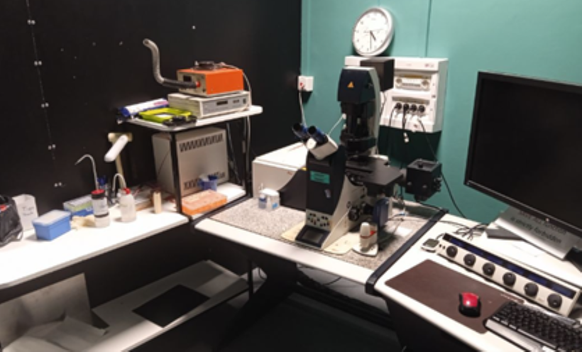
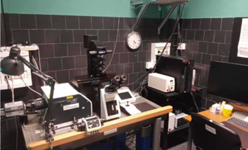
Leica TCS SP5 Confocal Microscope
The Leica TCS SP5 Confocal Microscope has 8 laser lines (405, 458, 476, 488, 496, 514, 543 and 633 nm) and 7 objectives (5x/0.15, 10x/0.3, 20x/0.7, 40x/0.75, 63x/1.4 OIL, 63x/1.2 W, 25x/0.95 W). AOBS (Acousto Optical Beam Splitter) is used to separate the excitation from emission light. Imaging method: scanning confocal, simulteanous 3 color detection using 3 spectral detectors plus transzmission mode.
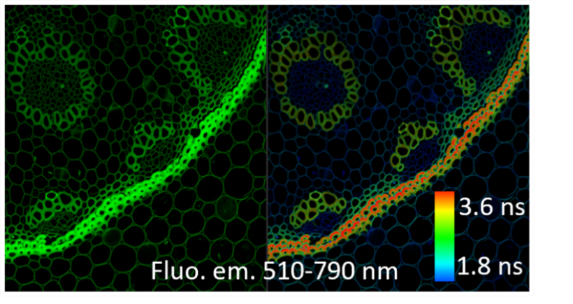
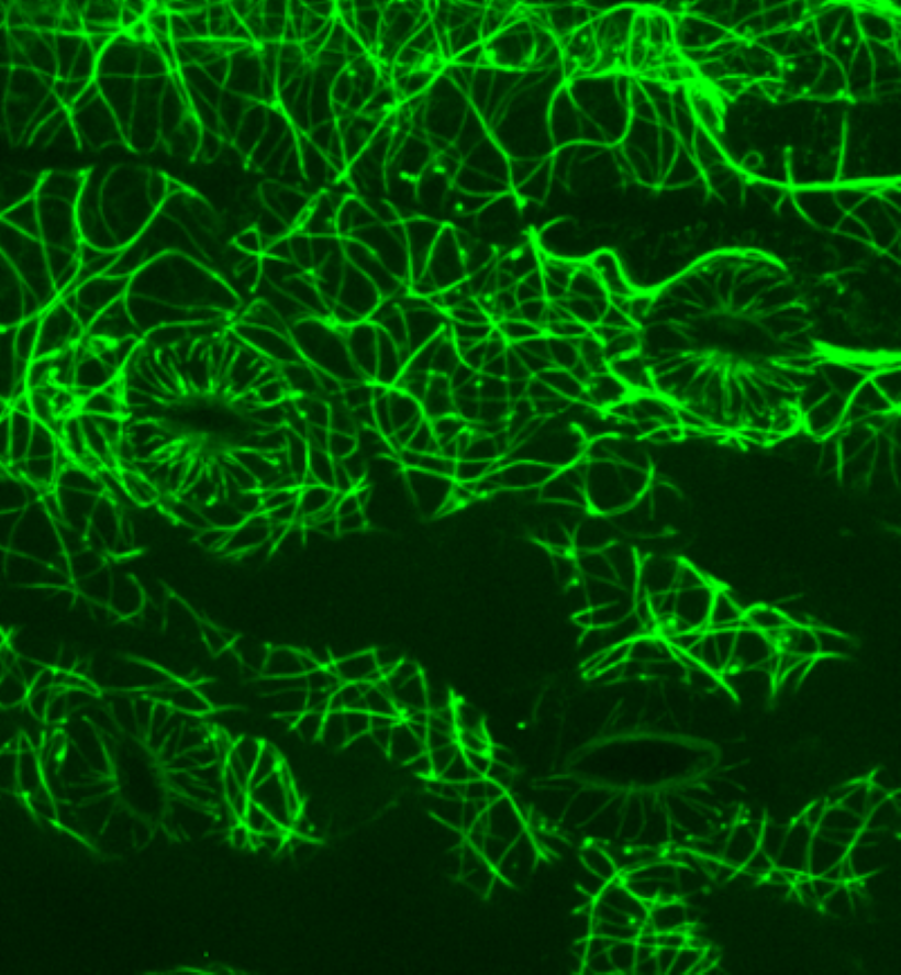
Microscopic photos by: Steinbach Gábor
Olympus FV1000 Confocal Microscope
The Olympus FV1000 Confocal Microscope has 6 laser lines (405, 458, 488, 515, 543 and 633 nm), and 6 objectives (10x/0.4, 20x/0.75, 40x/1.3 OIL, 60x/1.35 OIL, 40x/0.8 W and 60x/0.9 W). Available main dichroic beam splitters: BS20/80, DM405/488, DM/488/543/633, DM458/515 and DM 405/488/543. Imaging method: scanning confocal, stimultaneous 3 color detection, 2 independent spectral detectors + 1 additional PMT with emission filters + transmission mode.
VisiScope Spinning Disk Confocal Microscope
The VisiScope Spinning Disk Confocal Microscope has 4 lines (405, 488, 561 and 640 nm), and 6 objectives (4x/0.13, 10x/0.3, 20x/0.45, 40x/0.6, 60x/1.42 OIL and 100x/1.45 OIL). Detection is based on two Andor Zyla 4.2 PLUS cameras. High speed confocal imaging, simultaneous 2 color detection and sequential muntichannel acquisition available.
Microscopic photos by: Valkony Ildikó
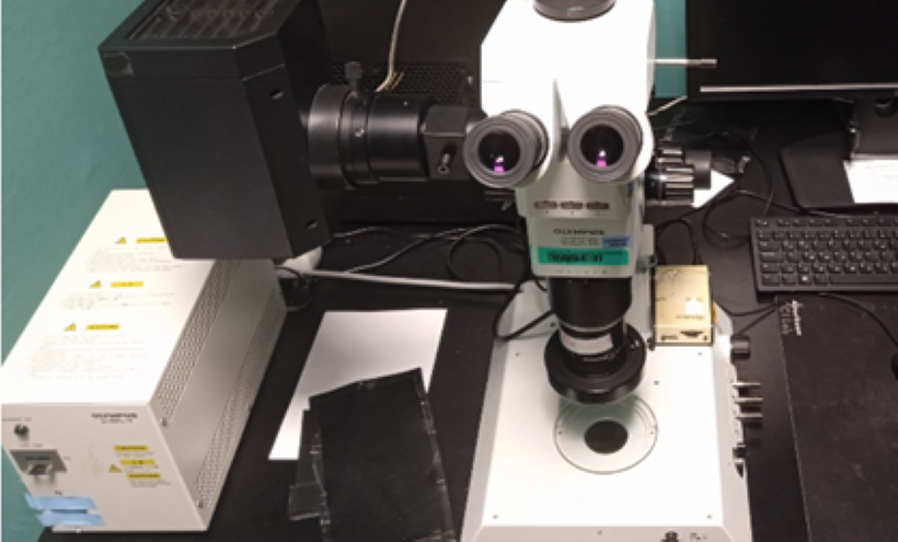
Olympus SZX12 Stereozoom Microscope
The Olympus SZX12 Stereozoom Microscope has halogen transmission (bottom), LED reflection (top) and Hg fluorescence (top) illumination. Imaging system: Cannon 200D camera. Available dichroic beam splitter cubes: DAPI, GFP/long pass, dsRed.
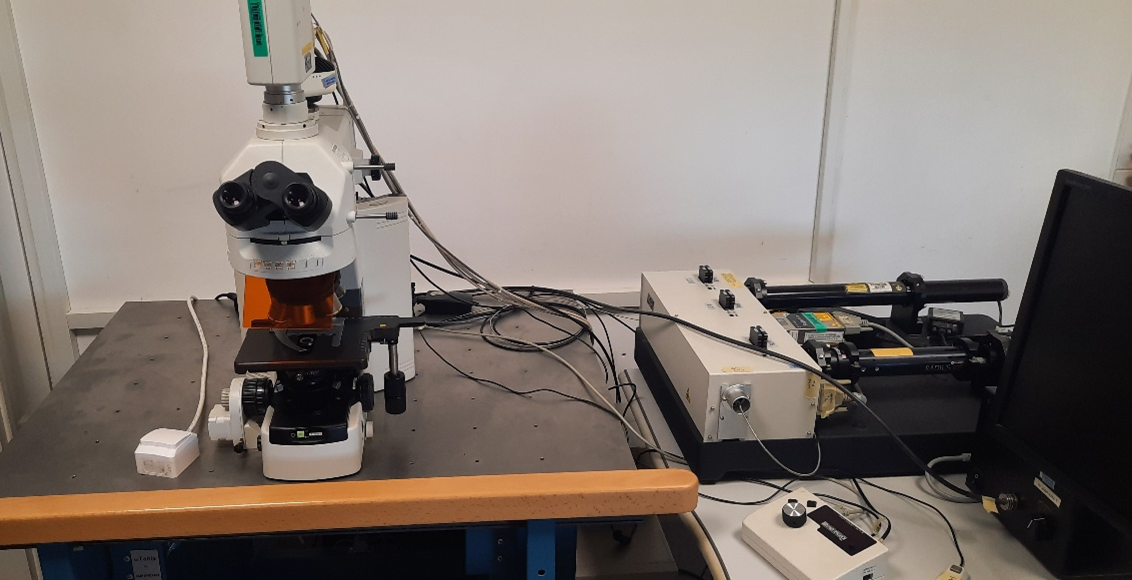
Nikon C1 Microscope
The Nikon C1 Microscope has 3 laser lines (405, 488 and 543 nm) and 6 objectives (4x/0.13, 10x/0.3, 20x/0.5, 40x/0.75, 60x/1.4 OIL and 100x/1.3 OIL). The microscope is suitable for bright field, fluorescence, and 3-channel confocal imaging.
Services
· 3D fluorescence imaging – confocal fluorescence multichannel high resolution image acquisition
· Live cell video / time-lapse imaging – incubation chamber with stabilized temperature and CO2 concentration
· Image processing and analysis – using ImageJ/FIJI and MATLAB, custom programs