 HUN-REN Biological Research Centre, Szeged
AFM/Raman laboratory
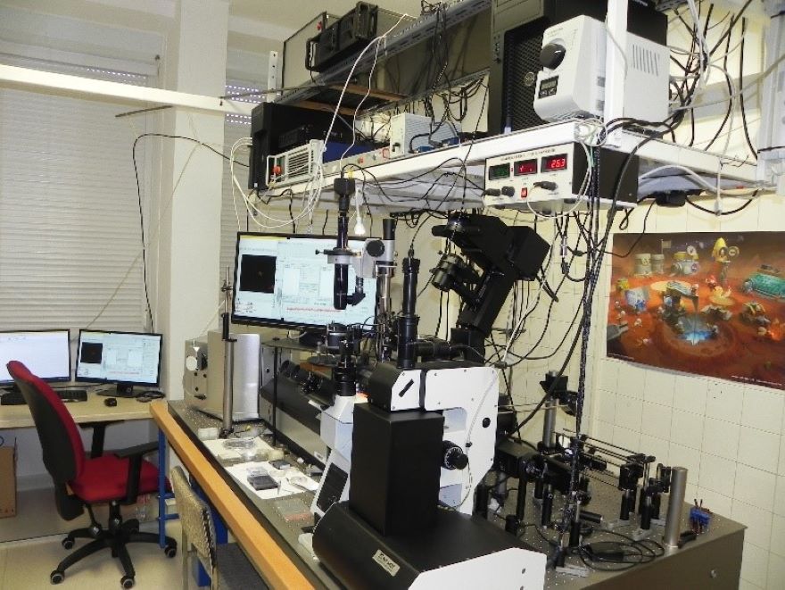
A scanning confocal Raman microscope built on an inverse optical microscope, integrated with an atomic force microscope (AFM), suitable for high-performance tip-enhanced Raman spectroscopy (TERS).
AFM: NT-MDT Ntegra II
szub-nanometer resolution imaging
pN resolution force measurements
high-frequency elacticity-mapping with imaging resolution
Raman confocal microscope: LSCM Spectra
Diffraction-limited, label-free chemical imaging based on inelastic light scattering (stage movement range: 70x70mm, piezo movement range: 100x100x10 microns)
Tip-enhanced, high-resolution, label-free chemical imaging (TERS)
Home-built optical tweezers expansion module
Cleanroom for microfabrication
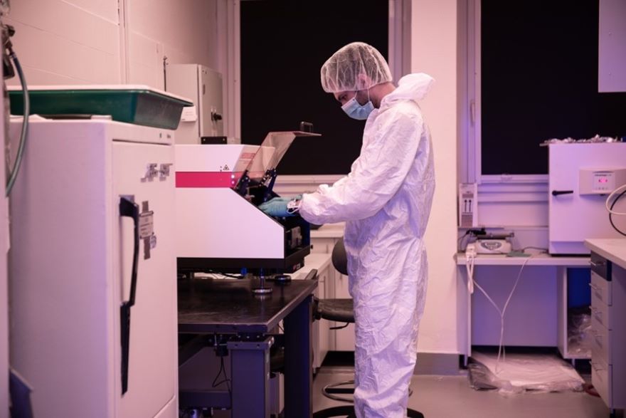
Dust-free (Class 100.000) laboratory for photo- and soft-lithography.
Heidelberg uPG101 micro pattern generator
0.6 micron minimum feature size
125X125 mm writing area
3 writing modes
Newport mask aligner (Newport 83210 Mask Alignment Fixture, Newport 68951 Digital Exposure Controller, Newport 4X4 500W NUV Illumination System)
For Cr-mask-based exposures
Hg- lamp with filter (for SU-8 exposure)
5"X5" mask size
Exposure time or dose can be set
Harrick plasma cleaner
Oxygen plasma treatments
30 W power
POLOS SPIN150i Spin coater
Maximum 6" substrate diameter
Maximum 12000 RPM spinning speed
Programmable
SPS 6800 Spin coater
Maximum 7" substrate diameter
Maximum 10000 RPM spinning speed
Programmable
Microfluidics workstation

Microscopy workstations tailored for microfluidics applications
Nikon TI-U epi-fluorescence microscopes with computer control
10X-100X objectives
several fluorescence filter cubes
phase-contrast imaging
motorized stage
laser-assisted autofocus
cage incubator with carbon dioxide
Syringe pumps
programmable
inject/withdraw
high precision microfluidic pump
Pressure controller
for driving flow in microfluidic chips
positive/negative pressure ( from -900 millibar to 6 bar)
4 channels
programmable
Scanning electron microscope
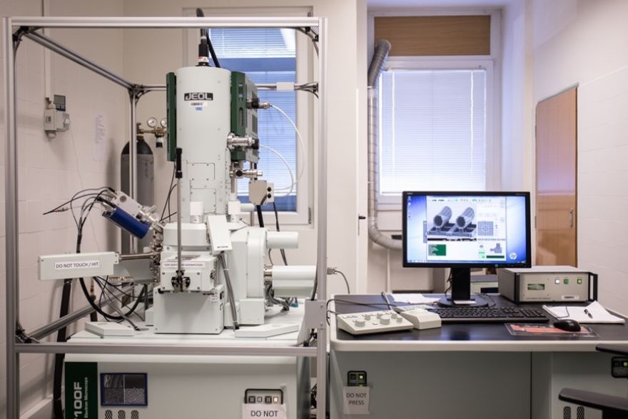
EOL JSM-7100F/LV electron microscope for morphological analysis. The instrument is able to operate in a low-vacuum mode for non-conducting/uncoated.
field-emission electron source
detectors: back-scattered and secondary electrons
resolution: 1.2 kV (30 kV), 3.0 nm (1 kV)
magnification: x 10 – x 1 000 000
accelerating voltage: 0.2 – 30 kV
sample movement: X: 70 mm, Y: 50 mm, Z: 40 mm, tilt: -5 - 70°, rotation: 360°
maximal sample size: 100 mm diameter, 40 mm height
equipped with magnetic noise-cancelling system
Low-vacuum (LV) mode
sample chamber pressure in LV mode: 10 Pa – 300 Pa
LV resolution: 1.8 nm (30 kV)
Sample preparation
critical point dryer for biological samples
Quorum Technologies K850
sputter deposition (Au, Cr)
Quorum Technologies Q150R
Two-photon polymerization laboratory
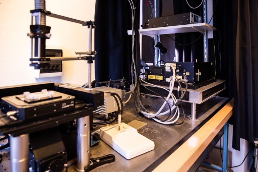
Laser polymerization workstation suitable for manufacturing micrometer-scale 3D polymer devices with sub-micrometer resolution. The device is suitable for parallel manufacturing.
Light source: Menlo C-Fiber 780A ultrashort pulse laser
wavelength: 785 nm
pulse length: 100 fs
pulse repetition frequency: 100 MHz
Sample positioning: Physik Instrumente P-563.3CL 3-axis piezo translator
maximal sample size: 300 mm x 300 mm x 300 mm, can be extended laterally
Minimum feature size: 200 nm
Parallelization: Holographic multiplication of the laser beam enables the simultaneous production of multiple copies of the polymerized structure.
Photopolimers at hand: SU-8, Ormocomp, Norland optical adhesive
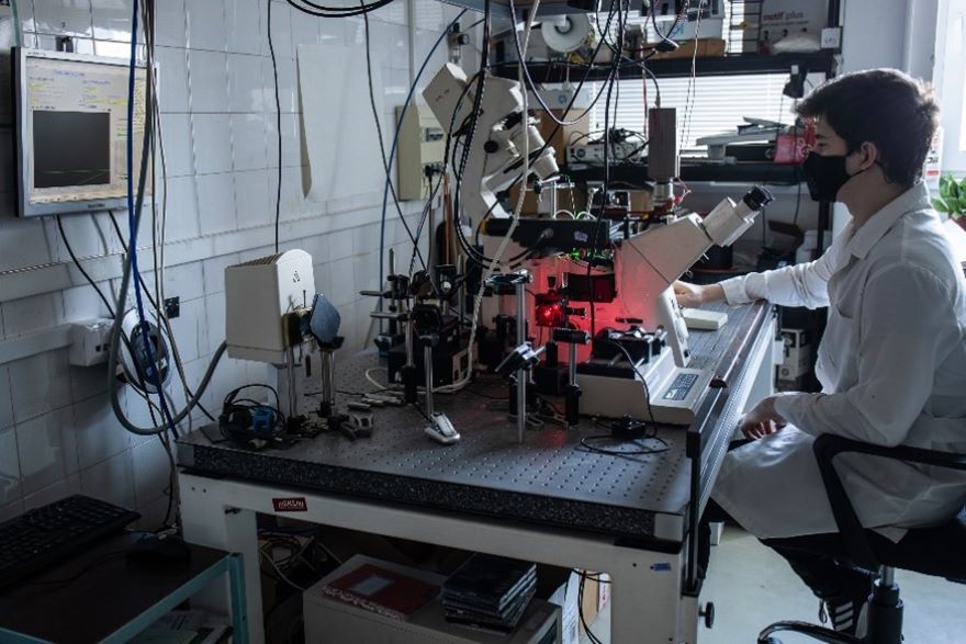
Holographic optical tweezers
Optical tweezers built onto a Nikon fluorescence microscope frame, creating multiple focal points for trapping simple and complex artificial structures or living cells.
63x, 1.2 NA water immersion objective
Holographic optical tweezers with ~200 mW effective trapping laser power
Hamamatsu ORCA Flash 4.0 high-sensitivity camera
Fluorescence filter sets
Toptica ICHROME laser source, 405 nm, 488 nm, 561 nm, 640 nm
Microscope stage with stepper motors and piezo translators
Heated sample holder
Laboratory of biological barrier models
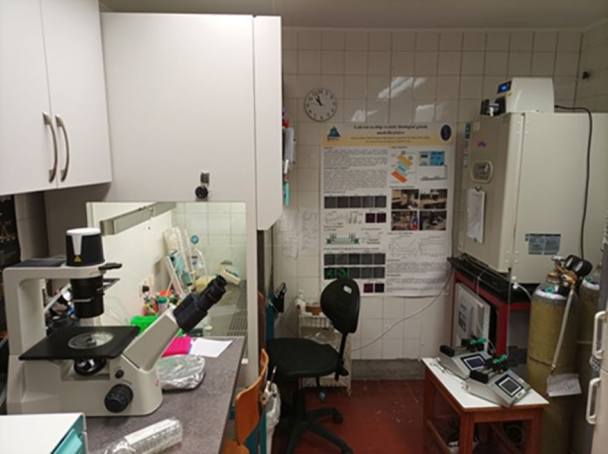
Instrument set for complex chip laboratory model-based testing of biological barrier systems (blood-brain barrier, intestinal epithelium, pulmonary epithelium, retinal barrier).
CO2-incubator (SANYO))
laminar flow hood (HERA Safe)
Phase-contrast microscope (Motic)
Zeta potential and dynamic light scattering measurements (Malvern Zetasizer Nano)
Broadband preamplifiers (voltage amplifier and current-voltage converter, Stanford Instruments)
Impedance spectroscopy (BioLogic – 0-1MHz, és HIOKI – 1MHz-600 MHz)
Integrated optics, electronics, and biosensorics laboratory
Instrumentation for testing biosensors created by combining optical, electrical, and microfluidic elements, and for studying the kinetics of light-induced biological processes
Microscopes (Zeiss Axiovert 200, Olympus IX71)
Syringe pumps (WIPI, kdScientific)
Motorized actuators (Marzhauser)
Lasers:
excimer laser (Lambda Physik Lextra 100)
N-YAG pulsed laser with optical parametric oscillator (Continuum)
He-Ne laser (Melles Griot, 632.8nm, 15 mW)
diode lasers (410-860nm, Roithner)
Fiber optics toolkit (ThorLabs)
Integrated optics and microelectrode-structures (Macz-Zehnder interferometers, dielectrophoresis-electrodes)
OWLS-instrumentation with a microradian precision rotating stage (Ealing
Electro Optics)
Flash-photolysis setup with optical multichannel analyzer (Andor iStar detector, Jobin-Yvon monochromator)
Roland film cutter
Sputterer (EmiTech)
Function generator (Tabor Electronics)
Digital oscilloscope (LeCroy Wave Runner)