 HUN-REN Biological Research Centre, Szeged
Jan 30, 2023 | News
The development of medicine and technology makes more and more tools and methods available for doctors and researchers to explore the molecular background of the disease progression and individual changes, knowledge of which enables personalized therapy. The first large-scale program to achieve these goals was the Human Genome Project (HGP, October 1990 - April 2003), which mapped the entire human genome. The following was the Human Cell Atlas (HCA), an international consortium founded in 2016 with more than 2,300 members that aims to create a reference map with the most detailed description of all cell types in the human body.
The research goals of Péter Horváth and his research group, the Lendület Laboratory of Microscopic Image Analysis and Machine Learning, are closely related to those defined by the HCA. In 2022, the group published their latest artificial intelligence (AI)-based single-cell image processing method in the prestigious journal Nature Biotechnology, taking cell characterization to a new level. Their method, called Deep Visual Proteomics (DVP), combines AI-based computer image analysis with laser microdissection microscopy and high-sensitivity mass spectrometry. The essence of DVP is that a microscope captures a high-resolution image of a paraffin-embedded tissue sample taken from the patient's tumor, from which a machine learning algorithm selects cells with abnormal morphology (computer-assisted microscopy isolation, CAMI), which are excised with a precision laser microscope and sent for molecular analysis. In the case of DVP technology, mass spectrometric analysis is performed where we can get a complete picture of the extracted single cell protein pool.
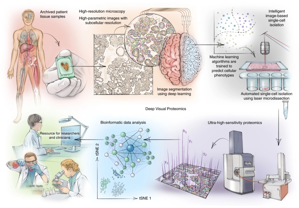
Deep Visual Proteomics workflow
With the development of the DVP method, scientists have at their disposal a technology that opens new doors in the world of image analysis. Taking advantage of the possibilities of this technology - with the support of the Chan Zuckerberg Foundation, the European Union's Human Cell Atlas projects and the Human Cell Atlas itself - Péter Horváth and his research team have established the Single Cell Centre, where they are looking for answers to biological basic research questions, such as how tumors develop, how they metastasize or what happens to cells during division.
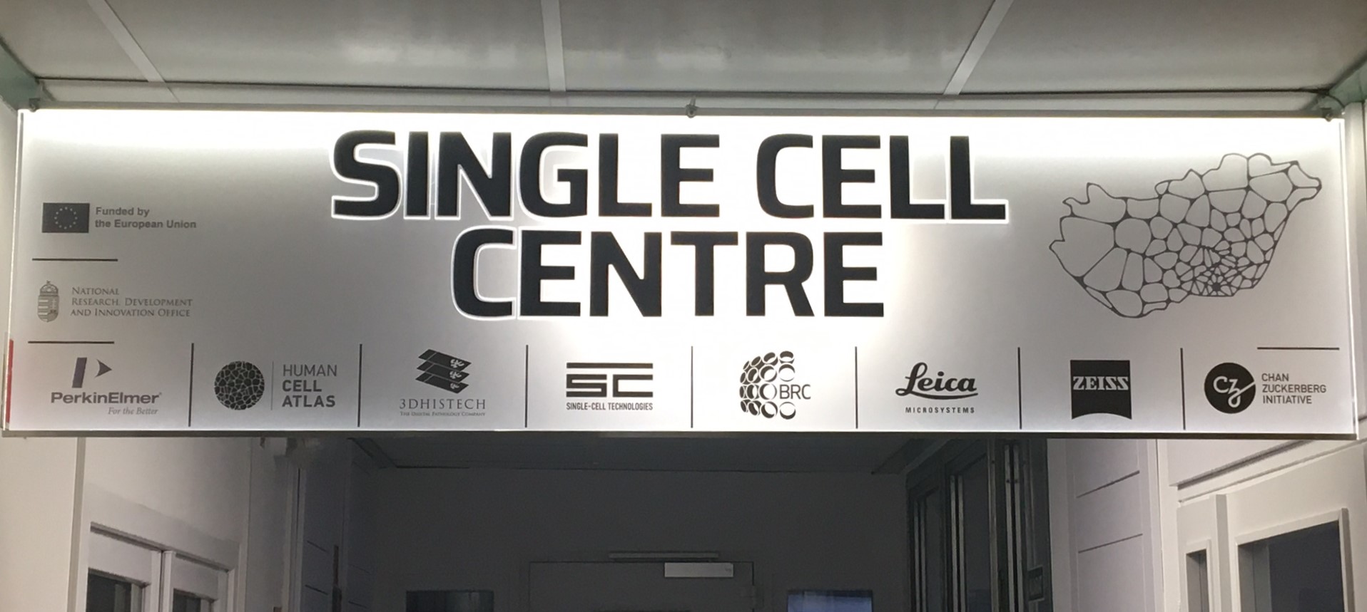
Business sign in the BRC Szeged
In order to achieve this, an automated system, unique in Hungary, has been set up, which performs every single step from the receipt of the sample to the extraction of the single cell in the Single Cell Centre. The professionalism is further enhanced by the fact that with the help of the barcode technology the entire history of the extracted single cell can be retrieved, starting from the patient's ID, through the applied experimental steps, up to the last working phase.
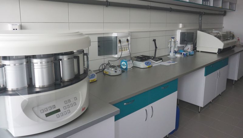
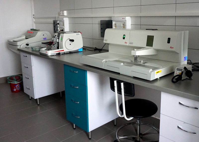
Leica tool park for processing tissue samples
Further molecular analyzes of the samples are performed with the help of cooperations with distinguished foreign institutes. Among the cooperation partners, the laboratories of Matthias Mann in Munich and Copenhagen are particularly noteworthy, with whom the group works very closely. Matthias Mann is one of the best experts in the field of proteomics, and his knowledge and guidance during the works are a great support. In addition, various omics studies are performed in Bergen, Lund, Zurich and Szeged in collaboration with other groups.
For the time being, the group is conducting research on cell lines (e.g. HeLa), but also wants to observe the changes in the cells on tumor tissue, especially on samples from patients with breast, lung and melanoma tumors. In this regard, thanks to international collaborations, there are already promising results in the study of tumor samples from patients. The study of a joint work with colleagues at the College of Helsinki was published, in which a sample from a patient with a pancreatic tumor was examined, but also a project collaboration with the Institute of Pathology and Molecular Pathology in Zurich, in which the genome and transcriptome analysis of cells from a rhabdoid renal tumor was recently completed.
As soon as the necessary permits are granted, artificial intelligence-based image analysis and single-cell extraction of tumor tissue samples can also begin at the Biological Research Centre, Szeged. A number of Hungarian grants are also available for the realization of the research goals, for example, the Thematic Excellence Program, which largely supported the establishment of the Single Cell Centre.
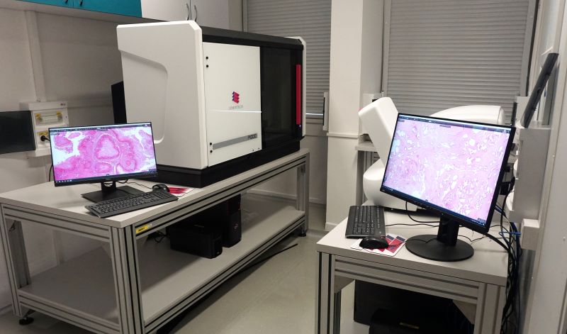
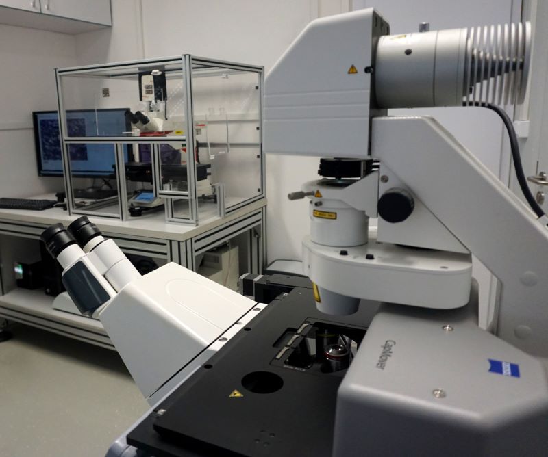
The first image shows 3DHistech P1000 and P250 slide scanners, and the second shows a Leica LMD6 and a Zeiss PALM laser microdissection microscope